Home
/ Plant Cell Under Microscope 40X Labeled - Powerpoint Lab Comparing Plant And Animal Cells : They are many different types of microscope that are used by.
Plant Cell Under Microscope 40X Labeled - Powerpoint Lab Comparing Plant And Animal Cells : They are many different types of microscope that are used by.
Plant Cell Under Microscope 40X Labeled - Powerpoint Lab Comparing Plant And Animal Cells : They are many different types of microscope that are used by.. The bulb of an onion is formed from modified leaves. Put a drop of water on the microscope slides. Unlike most plant cells, this species do not have a cell wall. See how a generalized structure of an animal cell and plant cell look with labeled diagrams. Under the microscope, plant cells are seen as large rectangular interlocking blocks.
Set up your microscope, place the onion root slide on the stage and focus on low (40x) power. 3) to draw and label a plant cell under 40x, a spider under 4x and human blood under 100x objective lens. Compare animal and plant cells and distinguish each type under the microscope. Do this step next to a drop of water, so the spores can be released into water. The cell wall is distinctly visible around each cell.
Https Plantenergy Edu Au Media Documents 8gmueuuebotlgey7x Original 8gmueuuebotlgey7x Pdf from Label the sketches to note the cell structures that you can identify. 2) to observe different cells/organisms using a virtual microscope. Diagram of plant cell under microscope. Make sure to give each figure: Cells walls should be visible: Examine larger specimens with the stereoscopic dissecting microscope. If you have questions regarding a microscope for viewing plant cells or plant stomata contact microscope world and we will be happy to help. Onion cells under a microscope requirements, preparation and observation.
The microscope is used for looking at many specimens that cannot be seen with the…
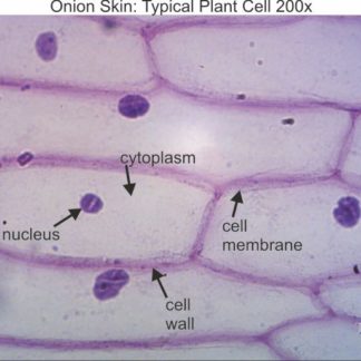
In this procedure, we will be using a conventional compound microscope. Plant cell under a microscope labeled labelled diagram of a plant cell under a microscope. Learn the structure of animal cell and plant cell under light microscope. This is the part used to look through the microscope. Experiment with different diaphragm apertures, as well as with different levels of focus and change the condenser lens setting (if your microscope can do this). Unlike most plant cells, this species do not have a cell wall. See how a generalized structure of an animal cell and plant cell look with labeled diagrams. The black sorus is riper than the white one. Tomatosphere tomatosphere specialized cells of the leaf syste. While photosynthesis takes place in the leaves of an onion containing chloroplast the little glucose that is produced from this process is converted in to starch starch granules and stored in the bulb. Animal cells results microscope observations *within the circles below, draw what you see. Whats people lookup in this blog: • label the magnification under which the plant cells are being observed (40x or 100x).
Experiment with different diaphragm apertures, as well as with different levels of focus and change the condenser lens setting (if your microscope can do this). Be sure to note any changes in the color, size, and shape of the cells. Do this step next to a drop of water, so the spores can be released into water. Even at low magnifications of, say, 10x to 40x, you will already see plenty of detail inside the cell. • label the magnification under which the plant cells are being observed (40x or 100x).
Typical Plant Cell 100x Dissection Connection from dissectionconnection.com.au Use tweezers to hold the frond, and use a dissecting needle to open sorus. The image below is a cross section of a plant captured under the richter optica hs1 student microscope at 400x magnification. Conclusions answer the following questions in your lab notebook. At 100x magnification you will be able to see 2mm. Can you identify any of the parts of the plant from the diagram shown above? Examine a variety of cells with the compound microscope and estimate cell size. At 400x magnification you will be able to see 0.45mm, or 450 microns. Label the cytoplasm and the cell membrane/wall (you won't be able to distinguish between the two).
Label the magnification under which the plant cells are being observed (40x or 100x).
• prepare sketches of your observed elodea cells under each set of conditions (aquarium water, 10% salt solution, and distilled water). Elodea are common freshwater aquarium plants. Switch to low power (10x). The compound microscope is a precision instrument. Set up your microscope, place the onion root slide on the stage and focus on low (40x) power. Images were taken on an inverted compound microscope using a 40x dic objective and. Experiment with different diaphragm apertures, as well as with different levels of focus and change the condenser lens setting (if your microscope can do this). Whats people lookup in this blog: Also indicate the estimated cell size in micrometers under your drawing. Unlike most plant cells, this species do not have a cell wall. While photosynthesis takes place in the leaves of an onion containing chloroplast the little glucose that is produced from this process is converted in to starch starch granules and stored in the bulb. At 400x magnification you will be able to see 0.45mm, or 450 microns. Can you identify any of the parts of the plant from the diagram shown above?
Set up your microscope, place the onion root slide on the stage and focus on low (40x) power. This slide was scanned using a 40x 080na objective. Images were taken on an inverted compound microscope using a 40x dic objective and. Make your sketches as accurate as possible. • label the magnification under which the plant cells are being observed (40x or 100x).
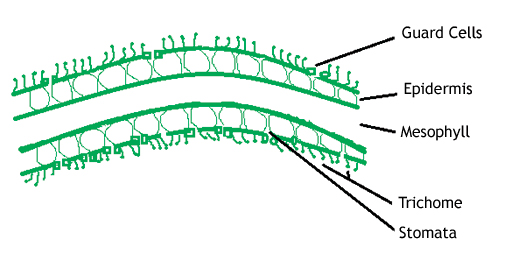
Plant Stomata Under The Microscope And What Stomata Tell You About Plant Habitat from www.microscopeworld.com Examine larger specimens with the stereoscopic dissecting microscope. Images were taken on an inverted compound microscope using a 40x dic objective and. Set up your microscope, place the onion root slide on the stage and focus on low (40x) power. In this procedure, we will be using a conventional compound microscope. Use tweezers to hold the frond, and use a dissecting needle to open sorus. Label the magnification under which the plant cells are being observed (40x or 100x). Examine a variety of cells with the compound microscope and estimate cell size. When carrying it, always use two hands, one on the base and one on the neck.
Identify the following structures on prepared slides and explain the functions of each:
How to observe cork cells under a microscope Machine vt recommended for you. This is the part used to look through the microscope. Learn the structure of animal cell and plant cell under light microscope. Even at low magnifications of, say, 10x to 40x, you will already see plenty of detail inside the cell. Observation of euglena under more powerful electron microscopes have revealed the presence of an ornamented pellicle under the plasma membrane. The black sorus is riper than the white one. Conclusions answer the following questions in your lab notebook. In this procedure, we will be using a conventional compound microscope. • label the magnification under which the plant cells are being observed (40x or 100x). At 40x magnification you will be able to see 5mm. Put on a coverslip, and ready to see. 3) to draw and label a plant cell under 40x, a spider under 4x and human blood under 100x objective lens.
To explore basic microscopy techniques objectives: plant cell microscope labeled. Microscopic video of an elodea leaf at three separate powers.
Share :
Post a Comment
for "Plant Cell Under Microscope 40X Labeled - Powerpoint Lab Comparing Plant And Animal Cells : They are many different types of microscope that are used by."

Post a Comment for "Plant Cell Under Microscope 40X Labeled - Powerpoint Lab Comparing Plant And Animal Cells : They are many different types of microscope that are used by."